Acute Exacerbation Of Copd Chest X Ray

1 initial symptoms of increased dyspnea increased sputum purulence and volume cannot be used to differentiate clinically between the two groups except with a chest radiograph.
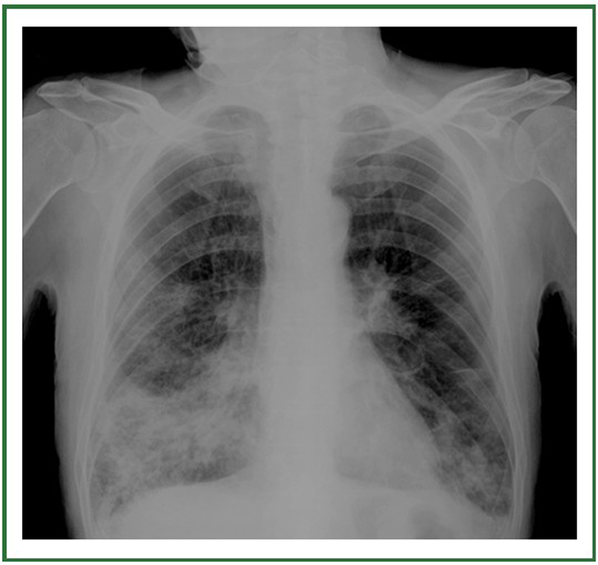
Acute exacerbation of copd chest x ray. 1 ucl respiratory university college london london uk nw3 2pf uk. It may be triggered by an infection with bacteria or viruses or by environmental pollutants. The resulting image may reveal enlarged lungs a flattened diaphragm or. 20 of chronic obstructive pulmonary disease copd patients admitted to hospital because of an exacerbation will have consolidation visible on a chest x ray.
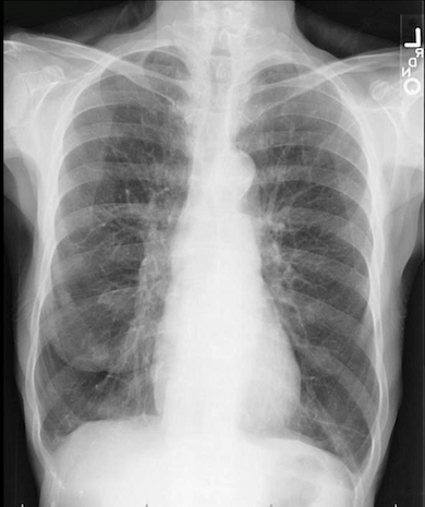
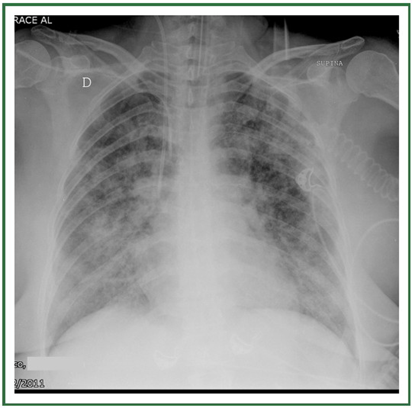
The chest x ray of a patient with acute copd exacerbation will show an increased anteroposterior diameter increased retrosternal airspace flattening of the diaphragm hyperinflation of the lungs bullae decreased lung markings a narrow vertical heart and the presence or absence of comorbidity. The authors take a stand that the presence of an infiltrate on a chest radiograph in a patient with copd should not be used to exclude them from a diagnosis of acute exacerbation of copd for two reasons. A chest x ray of someone with suspected chronic obstructive pulmonary disease or copd is a standard part of a diagnosis. And 2 the.
An x ray can also rule out other lung problems or heart failure. An acute exacerbation of chronic obstructive pulmonary disease or acute exacerbations of chronic bronchitis aecb is a sudden worsening of chronic obstructive pulmonary disease copd symptoms including shortness of breath quantity and color of phlegm that typically lasts for several days. If your doctor suspects you have chronic obstructive pulmonary disease copd you will be likely be asked to have a chest x ray. A chest x ray is a simple non invasive imaging technique that uses electromagnetic waves to create a one dimensional picture of your heart lungs and diaphragm.
Ct scans can also be used to screen for lung cancer. An exacerbation of copd periodic escalations of symptoms of cough dyspnea and sputum production is a major contributor to worsening lung function impairment in quality of life need for urgent care or hospitalization and cost of care in copd. Copd bullous emphysema bullous emphysema manifests on a chest x ray with areas of low density black with thinning of the pulmonary vessels predominantly affecting the upper zones the lower part of the lungs may appear denser whiter in normal subjects because of overlying breast tissue but in this individual the pulmonary vessels appear normal in this area. Contrast enhanced ct ct pulmonary angiography staging ct chest hrct chest etc.
A ct scan of your lungs can help detect emphysema and help determine if you might benefit from surgery for copd. Chronic bronchitis in chronic bronchitis bronchial wall thickening may be seen in addition to enlarged vessels. Findings of copd may be seen in a variety of ct chest studies e g. A chest x ray can show emphysema one of the main causes of copd.


